Kugako Sugimoto, NOST Tokyo
Originally published on the site of NL Agency
Summary
Masaki Ieda and his team created muscle cells by injecting three genes directly into the cardiac fibroblasts inside the mouse. They used a polycistronic virus vector that was able to carry three different genes and made the genes express simultaneously. The converting ratio from fibroblasts to muscle cells was doubled compared to the injection of each vector that contains one gene. This method is a quick and easy for regeneration of a heart by skipping time-consuming steps by transplanting of differentiated iPS/ES cells.
Masaki Ieda and his research team of the School of Medicine of the Keio University created muscle cells by injecting three genes into cardiac fibroblasts inside mouse’s body. This procedure is faster and easier compared to the creation of muscle cells from iPS or ES cells. Creating muscle cells from iPS and ES cells requires inducing, culturing in a lab, and transplanting differentiated cells to a damaged heart, which takes about one and a half months and skilled handling techniques. The technique of Ieda and his team will take about two weeks to induce muscle cells in a body.
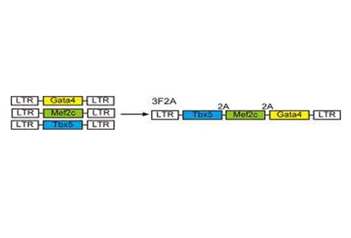
Creation of cardiac muscle cells from cardiac fibroblasts were already reported by U.S. research teams. Ieda and his team firstly used the polycistronic virus vector to introduce three genes to the cardiac fibroblasts for effective expression (Fig.1). Three genes lined up in a polycistronic virus vector and were expressed simultaneously in injected cells. The ratio of becoming muscle cells to fibroblasts was twice as much as by injecting three different kind of vectors each containing one type of gene. The team used genes, Gata4, Mef2c, and Tbx5 found as inducible genes of heart muscles by the Ieda’s team in 2010.
Fig.1 Polycistronic virus vector (School of Medicine, Keio University)
A human heart does not regenerate unlike a liver. Once the cardiac muscle cells are destroyed by myocardial infarction or dilated cardiomyopathy, cardiac fibroblasts increase, leading to cardiac fibrosis. In the worst case, a heart loses the function of pumping blood. Turning the cardiac fibroblasts into heart muscle cells regenerates the function of the heart.
The direct induction method is not only simple but also safe in comparison with the methods using iPS and ES cells. There is a concern about using iPS cells and ES cells in introducing unwanted cells such as cancer cells and undifferentiated cells to the body during transplant. Such a risk is very low for this direct method.
Surgery combining with a catheter and this direct induction method makes minimally invasive surgery possible if the take percentage of induced muscle cells becomes higher. It is also expected that this technique can be applied to creation of other types of organs in the future.
Source
Press release, School of Medicine, Keio University
iPS cells; Wikipedia
ES cells; Wikipedia
polycistronic ; MiMi
vector; Wikipedia
Gata4, Mef2c, Tbx5; Direct Reprogramming of Fibroblasts into Functional Cardiomyocytes by Defined Factors (2010); Cell, Volume 142, Issue 3, 375-386
myocardial infarction; Wikipedia
cardiomyopathy; Wikipedia
fibroblasts; Wikipedia


